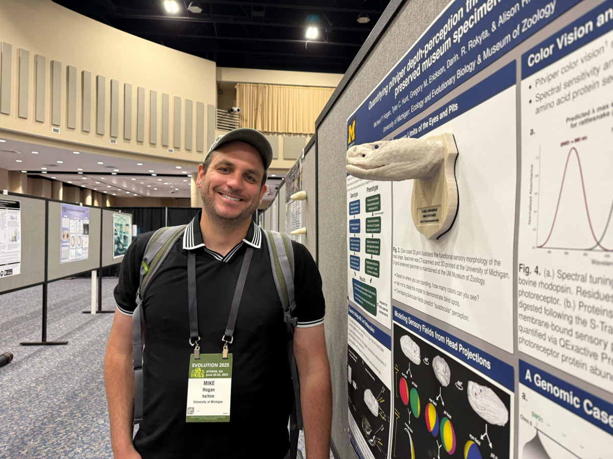
Thanks to the University of Michigan’s Museum of Zoology (UMMZ) Mike Hogan, in collaboration with Ramon Nageson and Alison Davis Rabosky, printed detailed 3D snakes using some of the Duderstadt Center’s 3D printers. Hogan, a postdoctoral research fellow at the University’s Department of Ecology and Evolutionary Biology, printed a total of six snakes. Hogan chose each of the six snake prints to bridge the work between his recent genetic PhD dissertation and his postdoctoral research work: the genetic expression of a snake’s thermal-sensing pit organ, and how it influenced the evolution of its venom and sensory systems.
Kathie Ferguson, the Manager of Fabrication Underground where the prints were made, enjoyed the project. It was printed on one of her favorite printers, the J850, as the machine can print different colors, flexibilities, and opacities all in one go. Hogan’s models utilized this technology for its eyes and thermal-sensing pit organ, each colored differently to more easily differentiate the unique features.
“[This project is] all done on the J850, because the printer has much flexibility,” Ferguson said with a smile. “They already printed one, but wanted two more. [The snake prints are] interesting unless you hate snakes, like my mother would be freaking out.”
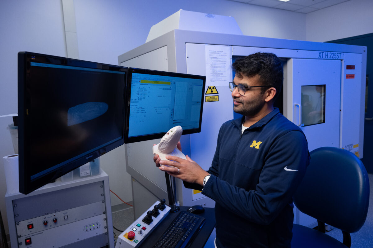
The first step to create these 3D replicas was to get computed tomography (CT) scans of the snakes he wanted to print. With the help of Ramon Nageson, a Research Specialist and the CT lab manager at the UMMZ, and the broad catalog of the 12,000 CT animal scans created by UMMZ, Hogan was easily able to get what he needed.
“The possibilities [for prints] are actually quite broad, because at this point, we have generated about 12,000 CT scans,” said Nagesan. “And it’s not just 12,000 snakes; it’s reptiles, it’s mammals, it’s fish, it’s birds, insects, as well as archaeological and paleontological objects. All of those could be printed, for various reasons and modalities.”
After being scanned, the specimen’s data goes through three more steps before printing. The last step involves getting the specimen pointed in a forward-facing direction. Unlike a human CT scan, many of these specimens are not scanned laying flat, but instead can be pointed every which way. Often, much of the time spent in the project is at this stage, rotating, aligning, and cropping the data to be printed.
“Our snakes could be upside down; they could be facing the corner,” Hogan said. “So when we get the CT data back, we have to expect that the specimen can be anywhere inside of this volume.”
With this handicap to churning out data quickly, Hogan wanted to come up with a solution to make the process faster. He realized that some of the snake’s facial features– like their eyes and prey thermal-sensing facial pits– could be used to anchor and align the specimen for analysis. With this discovery, Hogan created a code for technology to right the specimen on its own.
“In pursuit of my research on snake vision and their other sensory abilities, I realized some of the conserved head structures like the eyes and the thermal-sensing pits could be used to reliably ‘anchor’ or align the specimen for my analyses,” said Hogan. “I later realized this also sets up the data perfectly for 3D printing, which it turns out is a fantastic way to share my research findings.”
“I’m basically modeling the limitations of their visual field, but also looking at where these pits, these two holes, overlap,” Hogan said. “It turns out they have kind of a nice section in the middle where I’m calling their ‘quadocular overlap’ where you get to see all four colors. If you’re a mouse and you’re in that zone, you’re probably about to die, because their best depth perception is in that area.”
This code, which he hopes to release to the public this year, makes it easy for anyone to quickly align the data of the specimen to right the head. Hogan has even tried using his code on other vertebrates and has gotten similarly positive results.
“I’ve now tested it on a huge diversity of snakes,” Hogan said. “Some of them have weird appendages or a really long nose, and it doesn’t matter. It still works on any of those. I’ve even run it on salamanders and some other vertebrates that kind of have a similar head shape to the snakes.”
Another piece of information Hogan added to his 3D prints was adding unique printed number identifiers for each of the specimens, so that anyone could find out the exact location and history of that print. Alison Davis Rabosky, an associate professor in the Department of Ecology and Evolutionary Biology, a curator of reptiles and amphibians and director of UMMZ, mentioned the resonance of adding this new information.
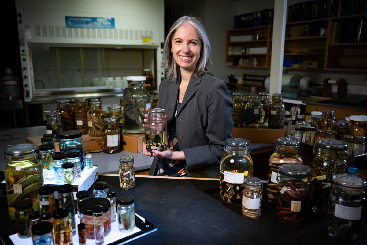
“One challenge we faced when doing some of these outreach events is that people lost contact or a sense of where the biodiversity data come from,” Davis Rabosky said. “And so, one of the things we tried to pair with these 3D prints is an activity we called the ‘story of the specimen.’ I would say everybody loved it. It connects [visitors] very, very directly to the data, and then they recognize there’s a scientific basis from which these 3D prints derive.”
These prints will continue to see the public in educational settings and even conferences. Though a part of the intention for these prints is primary and secondary education, there is learning for all ages with the use of the prints.
“I was at this meeting and I was having old-timer evolutionary biologists with canes coming over and getting really excited at these 3D prints,” Hogan said with a smile. “So, I think the range of the audience is pretty broad.”
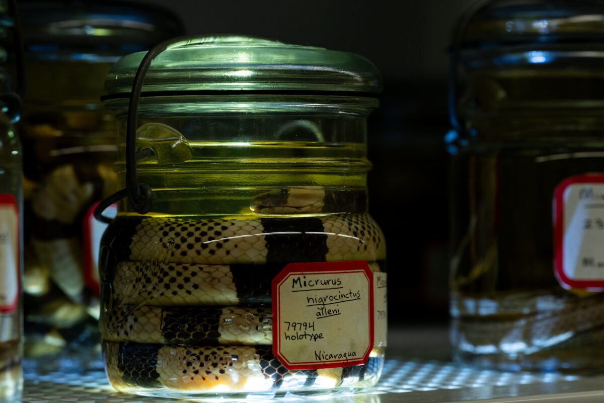
Not only is the range of impact broad, but so is the possible range of 3D prints. There are more than 500,000 preserved reptiles and amphibians, with 15 million zoological specimens total at the UMMZ. Nagesan hopes that there will only be more utilization of the creation of specimen prints in the future, as all have access to CT scan information.
“The number of which we have actually printed is quite small for the potential that exists,” Nagesan said. “And so, looking to the future, someone could come in and say, ‘actually, I want to look at different aspects of snakes,’ or not even snakes altogether, maybe: ‘I want to look at the hyoid bones of woodpeckers, where the tongue actually wraps around the skull.’ We have scans for that. Then, [if so desired, we can] print that. There’s a huge amount of potential here. We just scratched the surface.”
Much of UMMZ’s data is immediately accessible through the University’s Deep Blue Data website, or can be found here. The website has a series of collections divided by their collections division: mammals, herbs, fish, and birds, where the information is freely available to use in any way but commercially. Additionally, they invite readers to their SketchFab page, where more 3D models await their next use.
Photo Credits: Images courtesy of Mike Hogan and Michigan Photography
Article by Emma Powell